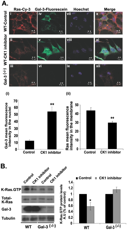
Figure 4. Inhibition of Gal-3 phosphorylation mediates K-Ras membrane mislocalization and downregulation.
A. The casein kinase inhibitor D4476 disrupts K-Ras localization in the cell membranes and attenuates export of Gal-3 from the nuclei of wt MEFs. Cells were plated on glass coverslips and treated for 24 h with D4476 or vehicle (control) as described in Methods. The cells were then fixed and labeled with Hoechst (purple), mouse anti-pan-Ras Ab followed by cy3-labeled donkey anti-mouse (red), and rat anti-Gal-3 Abs followed by fluorescein-labeled goat anti-rat Ab (green). Gal-3-/- MEFs served as a reference control. Upper panel: typical images, including triple-fluorescence merged images. Lower panel: (i) mean fluorescence intensity of fluorescein in the nuclei and (ii) of cy3 in the membrane (means ± SEM, n = 55 cells, **p<0.001). B. The casein kinase inhibitor CKI-7 reduces K-Ras.GTP expression in wt MEFs but not in Gal-3-/- MEFs. Cells from wt and from Gal-3-/- MEFs were grown in a 10-cm dish, treated for 24 h with 75 µM CKI-7 or vehicle (control), and then lysed. Lysates were subjected to quantification of active K-Ras.GTP, total K-Ras and Gal-3 by SDS–PAGE followed by immunoblotting with K-Ras and Gal-3 Abs as described in Methods. β-Tubulin served as a loading control. Left panel: typical immunoblots visualized by ECL. Right panel: levels of K-Ras.GTP expression. AU, arbitrary units; wt, wild type.

