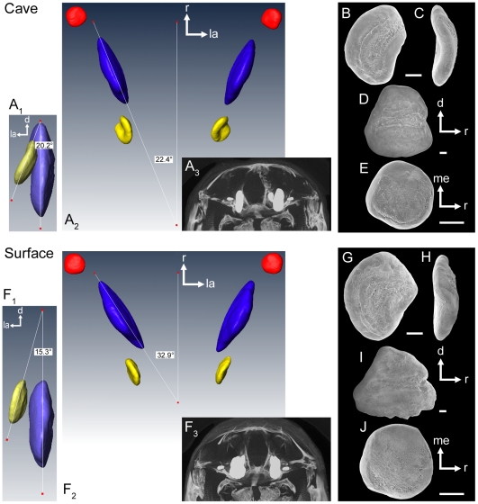
Figure 3. Position of otoliths in situ.
Three-dimensionally reconstructed otoliths from a cave (SL = 35 mm) and a surface fish (SL = 38 mm, both females) based on μ-CT analyses. In (A1) and (F1) the left asteriscus and sagitta are shown in caudal view. (A2) and (F2): dorsal view of left and right lapilli (red), sagittae (blue), and asterisci (yellow). (A3) and (F3): brightest point projection of μ-CT sections of the neurocranium and otoliths in situ shown in rostral view. (B–E) and (G–J): SEM images of the left otoliths of the cave and the surface fish in medial (B, D; G, I), rostral (C, H), and ventral views (E, J), respectively. B–C, G–H, asterisci; D, I, sagittae; E, J, lapilli. d, dorsal; la, lateral; me, medial; r, rostral. Scale bars = 100 µm.

