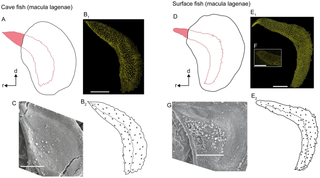
Figure 5. Orientation patterns of ciliary bundles on the macula lagenae.
(A) and (D), drawings of the asterisci and the overlying maculae lagenae showing the amount of the region of the sensory epithelium not overlain by the otolith in a cave fish (A, male; SL = 31 mm) and a surface fish from Río Oxolotán (D, female; SL = 34 mm). (B1) the left macula lagenae (mirrored) of the cave fish and (E1) the right macula lagenae of the surface fish of which the stereocilia of ciliary bundles were stained with TRITC-labelled phalloidin. The macula lagenae in the cave fish (B1) is only slightly bent while it is boomerang-shaped in the specimen from the surface habitat (E1). Note that the tip of the rostral arm in (E1) is slightly inflated. (F) shows the shape of an intact tip of the rostral arm of the macula lagenae of another surface fish (female; SL = 40 mm). (B2) and (E2), drawings of the same maculae as shown in (B1) and (E1), respectively, displaying a similar orientation pattern of the ciliary bundles in cave (B2) and surface fish (E2). (C) and (G), SEM images of the right macula lagenae of another cave fish (male; SL = 30 mm) and of the left macula lagenae (mirrored) of another surface fish (female; SL = 40 mm). d, dorsal, r, rostral. Scale bars = 100 µm.

