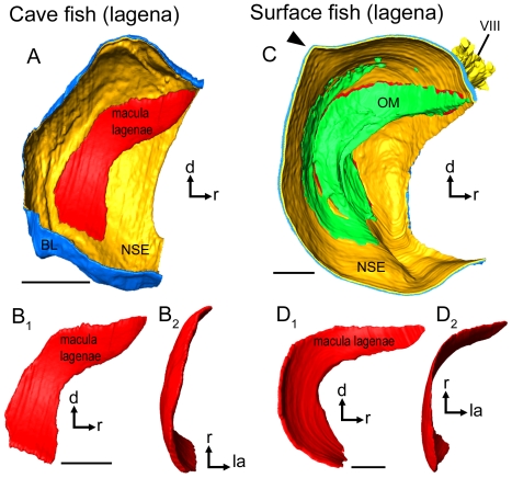
Figure 6. Three-dimensionally reconstructed lagena and macula lagenae of cave fish and surface fish from Tampico.
(A) and (C), lagenae in lateral view showing the macula, (the otolithic membrane), the non-sensory epithelium, and the basal lamina in a cave (A; male, SL = 35 mm) and a surface fish (C; female, SL = 52 mm). Note that the lagena in the surface fish (C) displays a distinct caudo-dorsal ‘edge’ (black arrowhead), while the lagena of the cave fish lacks this “edge” (A). This feature corresponds to the prominent posterodorsal edge of asterisci from surface fish (see Figure 5D vs. A). (B) and (D) display differences in curvature and especially shape of the macula lagenae of cave (B) and surface fish (D). The maculae lagenae are shown in lateral (B1, D1) and dorsal view (B2, D2). BL, basal lamina; d, dorsal; NSE, non-sensory epithelium; la, lateral; OM, otolithic membrane; r, rostral; VIII, part of the eighth cranial nerve innervating the lagena. Scale bars = 100 µm.

