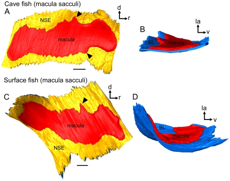
Figure 9. Three-dimensionally reconstructed macula sacculi of cave and surface fish (Tampico).
(A) and (C), maculae sacculi in lateral view showing the macula and the non-sensory epithelium. Black arrowheads label the ventral and dorsal bulges of the macula. (B) and (D), maculae sacculi in caudal view displaying the different amount of three-dimensional curvature. The strong curvature of the macula sacculi of the surface fish correlates with a thick sagitta whereas the flat almost two-dimensional macula comes along with a rather flat sagitta of the cave fish specimen. BL, basal lamina; d, dorsal; la, lateral; NSE, non-sensory epithelium; r, rostral; v, ventral. Scale bars = 100 µm.

