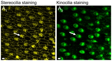
Figure 11. Confocal images of double labeling of a saccular epithelium.
(A1), stereocilia of ciliary bundles stained with TRITC-labeled phalloidin. (A2), kinocilia of the same ciliary bundles stained with anti-bovine α-tubulin mouse monoclonal antibodies and Alexa Fluor 488 conjugated anti-mouse secondary antibodies. White arrows indicate the orientation of ciliary bundles based on the position of the kinocilium. Scale bars = 2 µm.

