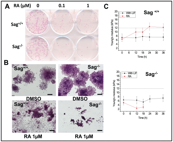
Figure 2. Sag deletion blocks RA-induced differentiation.
Sag+/+ and Sag−/− mES cells were seeded in 6-well plate and treated with RA at indicated concentrations for 6 days. Colonies were stained with AP (A) and counter-stained with hematoxylin (B), followed by photography. Bar size = 50 nm. Cells were seeded in geletin-coated glass coverslips and cellular stiffness was measured by AFM nanomechanical analysis at 0, 12, 18, 24 and 36 hrs post RA (1 µM) treatment. Shown is x ± SD from three independent experiments (C).

