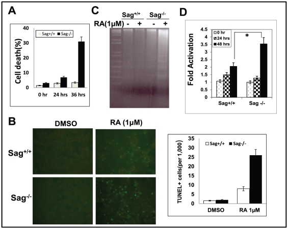
Figure 3. Sag deletion sensitizes mES cells to RA-induced apoptosis.
mES cells with genotype of Sag+/+ and Sag −/− were treated with DMSO control or 1 µM RA for indicated time periods, followed by trypan blue staining (A), TUNEL staining at 36 hrs (B, left panel), with quantification of TUNEL positive cells graphed (B, right panel), DNA fragmentation assay at 36 hrs (C), and caspase-3 activity assay at the indicated time point (D). *, p<0.05.

