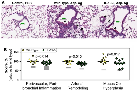
Figure 7. Histological changes in the lungs of wild type and IL-19-/- mice primed and challenged with Aspergillus fumigatus antigen.
Wild type and IL-19-/- mice were of the BALB/c background strain. Priming and challenge with Aspergillus antigen was as indicated in Figure S1. Control animals were given saline (PBS) intranasally. (A) Photomicrographs of lung sections from wild type or IL-19-/- mice. The sections were stained with periodic acid schiff and digitally scanned. The software was used to generate the scale bar (50 µm). Perivascular/peribronchial inflammation (arrows), pulmonary arterial remodeling (stars) and mucus cell hyperplasia (arrow heads) are indicated. (B) Scores for perivascular/peribronchial inflammation, arterial remodeling and mucus cell hyperplasia in the lungs of wild-type (circles) or IL-19-/- (diamonds) mice. The gray boxes outline the values from the groups of control animals depicting the scores spanning the 25% and the 75% quartiles. Dots represent individual data from Aspergillus antigen primed and challenged mice. Horizontal lines show medians. Three independent experiments were performed during a time span of 6 years. Therefore, scores needed to be standardized. For this reason, scores were plotted relative to the respective median scores of primed and antigen-challenged wild-type mice for each experiment and then the data were pooled. Significance levels were calculated with the Mann-Whitney-U test.

