
Fig 1.
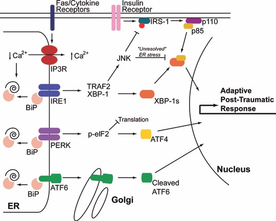
ER stress pathways activated post-burn are similar to those mediating type 2 diabetes. Burn injury causes multi-organ ER stress characterized by activation of all three arms of the ER stress response. In animal burn models, hepatic ER calcium stores are depleted most likely by activation of IP3R calcium channels leading to accumulation of unfolded polypeptides and activation of IRE1, PERK and ATF6 [53]. The mechanism by which the IP3R is activated is still unclear, but may be mediated by Fas receptor activation or calcium mobilizing cytokines (see text for details). Activation of JNK is prominent post-burn, leading to IRS-1 phosphorylation and suppression of PI3K activation (schematically represented by the p110 and p85 subunits) and ultimately insulin resistance [54, 55]. Insulin resistance in burn patients persists for an extended period of time (‘unresolved’ ER stress) similar to type 2 diabetes [78]. This may be mediated by suppression of XBP-1s/p85 transcriptional activity by JNK [60].