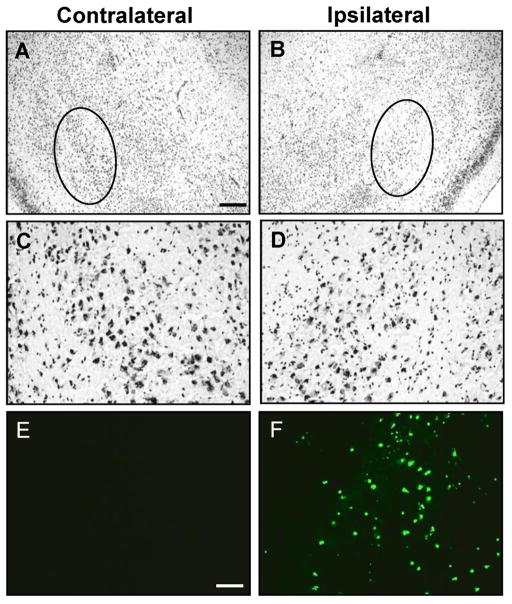
Figure 2.
Intraamygdaloid KA injection induces acute cell death within the ipsilateral amygdala. A–D, Representative images of Nissl staining in the contra- (A,C) and ipsilateral (B,D) amygdala 24 hours after intraamygdaloid KA injection. A,B, Disrupted Nissl staining in the ipsilateral amygdala (B, circle) and the corresponding unaffected contralateral region (A, circle). C,D, Higher magnification images of the Nissl stained regions demarcated by circles in panels A and B, respectively. E,F, TUNEL labeling of sections adjacent to panels A and B. Scale bars: 300 μm (AD), 75 μm (E–F).

