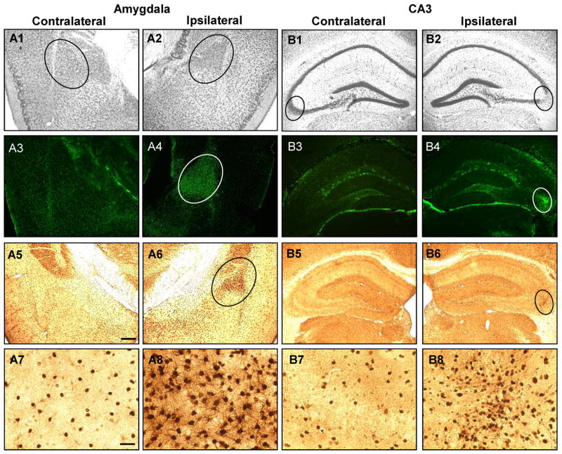
Figure 4.
Intraamygdaloid KA injection causes chronic injury associated with astrogliosis and ADK overexpression in the ipsilateral amygdala and CA3. A1,2;B1,2, Representative Nissl staining of the injured (A2, B2; circles) and corresponding non-injured (A1, B1; circles) amygdala (A1,2) and CA3 (B1,2) 3 weeks after KA injection. A3-8;B3-8, Representative images of GFAP (A3,4;B3,4) and ADK (A5-8;B5-8) immunoreactive material in the amygdala (A3-8) and CA3 (B3-8). Increased GFAP (A4,B4; circles) and ADK (A6,B6; circles) immunoreactive material is only present in the ipsilateral injury site and correspond to disrupted Nissl staining. A7,8;B7,8, High magnification images obtained from the contralateral amygdala (A7) and CA3 (B7) and within the area demarcated by circles in panels A6 and B6 for the ipsilateral amygdala (A8) and CA3 (B8), respectively. Scale bars: 300 μm (A1-6;B1-6), 37.5 μm (A7,8;B7,8).

