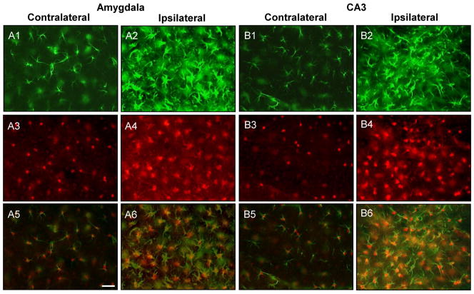
Figure 6.
ADK upregulation colocalizes with astrogliosis in the injured amygdala and CA3. A1-4;B1-4, Representative images of GFAP (A1,2;B1,2, green) and ADK (A3,4;B3,4, red) double labeled sections that were acquired from within the amygdala and CA3 three weeks after intraamygdaloid KA injection. The ipsilateral amygdala (A2,4) and CA3 (B2,4) have increased GFAP (green) and ADK (red) immunoreactive material compared to the contralateral hemispheres (A1,3;B1,3). A5,6;B5,6, Merged fluorescence image of GFAP and ADK immunoreactive material. The increased levels of ADK immunoreactive material (red) colocalizes with reactive astrocytes (GFAP, green) within the ipsilateral amygdala (A6) and CA3 (B6). Scale bars: 37.5 μm (A1-6;B1-6).

