
Figure 6. IL-12p40 expression is exaggerated in TLR2 KO microglia following exposure to intact S. aureus and the TLR9 ligand ODN.
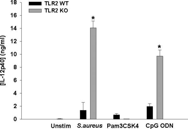
Primary TLR2 WT and KO microglia were seeded at 2 × 10⁵ cells per well in 96-well plates and incubated overnight. After 24 h, cells were exposed to heat-inactivated S. aureus (107 cfu/well), Pam3CSK4 (1 μg/ml), or CpG ODN (0.1 μM) for 24 h, whereupon IL-12p40 production was quantitated by ELISA. Significant differences between TLR2 KO versus WT microglia are indicated by asterisks (*, p < 0.001). Results are reported as the mean ± SD of three independent wells for each experimental treatment and were identical across three separate experiments.