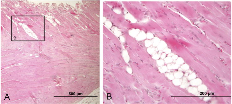
Figure 1.
(A) A mouse supraspinatus muscle at 8 weeks following tenotomy of both the supraspinatus and infraspinatus tendons shows several clusters of adipocytes within the muscle. (B) Adipocytes with the characteristic signet ring shape are clearly seen in a magnified view of the outlined area in (A). The muscle fibers show atrophy and degeneration, as evidenced by the small fiber size, centralized myonuclei, and rouleaux formation of myonuclei. [10X objective used in (A) and 40X objective used in (B); H & E stain.]

