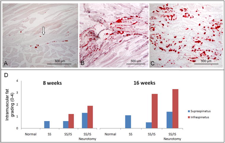
Figure 3.
(A) A normal rat supraspinatus muscle stained with Oil red O showing very few intramuscular fat deposits and intramyocellular fat droplets. The supraspinatus tendon can be seen at the center of the muscle (arrow) and the muscle fibers can be seen above and below the tendon. (B) The infraspinatus muscle of a rat 16 weeks following tenotomy of the supraspinatus and infraspinatus tendons. There are high numbers of fat deposits (seen as red dots). (C) The infraspinatus muscle of a rat 16 weeks following tenotomy plus neurotomy showing high levels of intramuscular fat. [10X objective; Oil red O stain.] (D) Histology grading results are shown for intramuscular fat on Oil red O stained histology sections. Normal muscles showed no fat. After tenotomy of the supraspinatus and infraspinatus tendons, the infraspinatus muscle had more intramuscular fat than the supraspinatus muscle. The 16-week specimens had more intramuscular fat than the 8-week specimens within each group.

