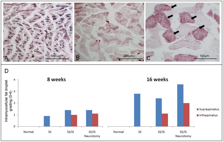
Figure 4.
(A) The supraspinatus muscle of a rat at 16 weeks following tenotomy of the supraspinatus tendon showed a number of muscle fibers stained with Oil red O. However, there was almost no intramuscular fat observed. (B) A micrograph of the supraspinatus muscle of a rat at 16 weeks following tenotomy of the supraspinatus and infraspinatus shows higher levels of intramyocellular fat droplets in the muscle fibers. (C) A higher magnification of (B) shows fine, granule-shaped fat droplets within muscle fibers. A subset of muscle fibers showed high levels of fat droplets (arrows), making them appear as darkly stained fibers at low magnification. [10X objective used in (A), 20X objective used in (B), and 40X objective used in (C); Oil red O stain.] (D) Histology grading for intramyocellular fat droplets on Oil red O stained sections is shown. The normal muscles showed no fat droplets while the muscle that had received tenotomy only or tenotomy plus neurotomy showed intramyocellular fat droplets. The supraspinatus muscle had more fat droplets than the infraspinatus muscle when there had been tenotomy of both tendons. Overall, the 16-week specimens showed more fat droplets than the 8-week specimens.

