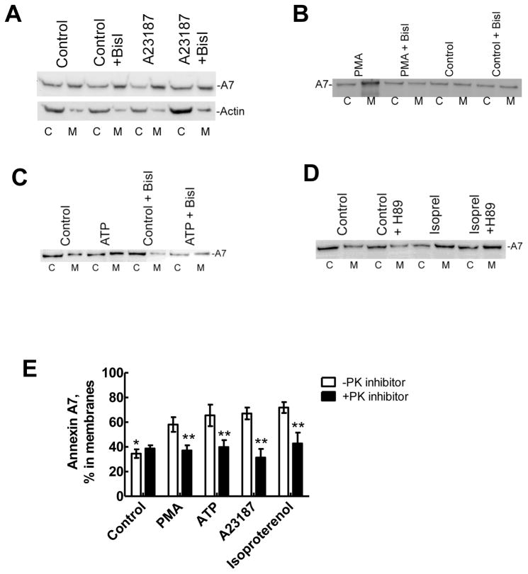
Figure 3. Membrane-association of annexin A7 is inhibited by protein kinase inhibitors.
Alveolar type II cells were incubated for 30min without or with protein kinase inhibitors, bisindolylmaleimideI (BisI) or H-89 before treatment for 30min without or with indicated secretagogues. Equal proportions of the membrane (M) and cytosol (c) fractions were probed for the annexin A7 levels by western blot analysis. Western blot for fractions from cells treated with A23187 (A) shows increased membrane-association of A7, which was inhibited in the presence of BisI. The distribution of actin was not affected by either A23187 or BisI. Similarly, PMA (B), ATP (C) and isoproterenol (D) increased membrane-association of A7, which was inhibited by respective protein kinase inhibitor. E. Results of densitometry analysis of western blot images from all experiments. Results are relative distributions in the membrane fraction as a percent of the total and are mean ± SE of 3 –12 cell preparations. * P < 0.05 in comparison to all other conditions in the absence of protein kinase (PK) inhibitor, ** P < 0.05 in comparison to respective condition in the absence of PK inhibitor (BisI or H-89, as appropriate).

