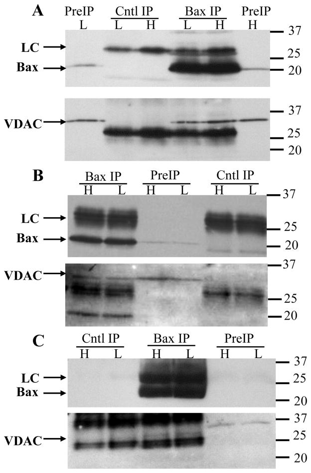
Figure 1. VDAC1 co-immunoprecipitates with Bax.
Granule neurons were maintained in high or low K for 5h then solubilized with 1% digitonin. The extracts were immunoprecipitated with the 1D1 antibody, or an irrelevant mouse monoclonal (A, C) or goat polyclonal (B) antibody (as control), separated by SDS-PAGE, blotted to PVDF membranes, then probed for Bax (top image of each pair), stripped, and re-probed for VDAC1. Three independent experiments are shown in A–C to demonstrate the variable recovery of VDAC1. (A) High VDAC1 recovery with 14 million neurons solubilized with 700μl. Protein concentrations in the high K control and 1D1 extracts were 557 and 547 μg/ml, respectively, and in the low K control and 1D1 extracts were 551 and 567 μg/ml, respectively. Prior to immunoprecipitation, the samples were concentrated to final protein concentrations of 1331, 1459, 1460, and 1450 μg/ml, respectively. (B) Moderate VDAC1 recovery with 6 million neurons solubilized with 525 μl. Protein concentrations were not determined in this experiment. The extracts were not concentrated prior to immunoprecipitation. (C) Weak VDAC1 recovery with 10.5 million neurons solubilized with 525 μl. Protein concentrations in the high K control and 1D1 extracts were 769 and 717 μg/ml, respectively, and in the low K control and 1D1 extracts were 491 and 495 μg/ml, respectively. Prior to immunoprecipitation, the samples were concentrated to final protein concentrations of 1986, 1930, 1235, and 1319 μg/ml, respectively. Occasionally, as in image C, the secondary antibody did not bind the control antibody light chain as strongly as the 1D1 light chain (although this was not true for the heavy chain; not shown), which resulted in undetectable light chains with short exposure times. The absence of Bax in the preIP lanes is also due to the short exposure time. Longer exposures confirmed the presence of the light chains and preIP Bax (not shown). Both Bax and VDAC1 in the initial extracts occasionally migrated more slowly than in the immunoprecipitates, which was attributed to interference by excess loading of digitonin. Molecular weight markers are indicated to the right of the blots. H: high K; L: low K; PreIP: initial digitonin extract; Cntl IP: control immunoprecipitation; Bax IP: Bax 1D1 immunoprecipitation; LC: antibody light chain.

