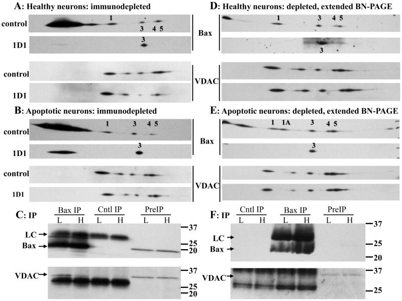
Figure 4. Analysis of Bax and VDAC1 oligomers following 1D1 Bax immunodepletion.
(A, B) Ten million neurons maintained in high (A) or low (B) K for 5h were solubilized with 525μl 1% digitonin. Protein concentrations in the high K control and 1D1 extracts were 438 and 521 μg/ml, respectively, and in the low K control and 1D1 extracts were 367 and 423 μg/ml, respectively. Prior to immunoprecipitation, the samples were concentrated to final protein concentrations of 993, 1403, 982, and 944 μg/ml, respectively. The extracts were immunodepleted of Bax overnight with the 1D1 antibody or an irrelevant mouse monoclonal antibody as control (top image of each pair). Aliquots (25μg protein) were subjected to BN-SDS PAGE, and immunoblotted for Bax and VDAC1 using the 1D1 and N18 antibodies, respectively. Bax oligomer 3 resisted immunoprecipitation. Note that oligomer 1A is not detectable in B. (C) Analysis of the immunoprecipitates from A and B for Bax and VDAC1, demonstrating strong recovery of VDAC1. (D, E) Seven million neurons maintained in high (D) or low (E) K were solubilized with 350 μl 1% digitonin. Protein concentrations in the high K control and 1D1 extracts were 698 and 641 μg/ml, respectively, and in the low K control and 1D1 extracts were 595 and 691 μg/ml, respectively. Prior to immunoprecipitation, the samples were concentrated to final protein concentrations of 1664, 1529, 1430, and 1612 μg/ml, respectively. The depleted samples were processed as above except that 36 μg protein was loaded, and the BN PAGE was performed for 240 min rather than 120 min to increase separation of the oligomers (with one consequence being that most of the monomer had migrated off the bottom of the gel). No change in VDAC1 oligomer intensities were observed (see Table 3). Representative images from 2 of 4 independent experiments are shown. (F) Analysis of the immunoprecipitates from D and E for Bax and VDAC, demonstrating weak recovery of VDAC1. As in Fig. 1, the control antibody light chain and preIP Bax are not detectable with the short exposure time. H: high K; L: low K; PreIP: initial digitonin extract; Cntl IP: control immunoprecipitation; Bax IP: Bax 1D1 immunoprecipitation; LC: antibody light chain.

