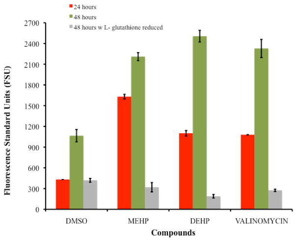
Figure 2.

Comparison of changes in the mitochondrial membrane potential among TK6 cells exposed for 24 and 48 hours to the IC50's of DEHP (234μM) and MEHP (196μM). For the 48 hours exposure assay the average level of mitochondrial membrane permeability caused by DEHP and MEHP presented P-values of 0.002 and 0.024, respectively when compared with the negative control. Also no significant difference was observed among the positive control (valinomycin) with MEHP and DEHP (P-value >0.10). Comparison of the changes in the mitochondrial membrane potential generated on TK6 cells exposed for 48 hours to the IC50's of DEHP (234μM) and MEHP (196μM) with 500μM of L- glutathione is also presented an illustrates the reduction effect.
