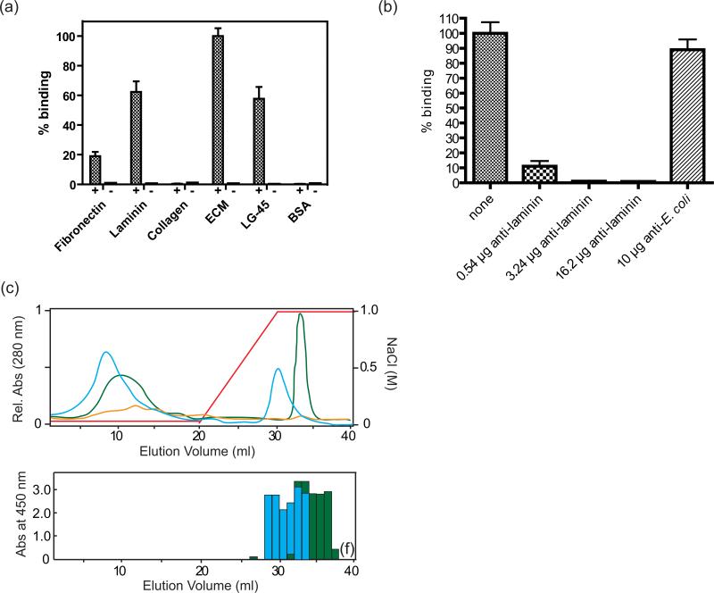
Figure 4. Laminin binding activity of Ail.
(a) Binding assay for Ail-expressing bacteria to human ECM proteins and LG4-5, a fragment of laminin. Numbers of bound bacteria to laminin, fibronectin, collagen, ECM, LG4-5, and BSA are shown as bars. Standard deviations are shown by error bars. Controls contain cells expressing empty vector. Lanes labels with + contain Ail, while lanes labled with – are controls. (b) Anti-laminin can block binding to ECM by Ail-expressing cells. ECM-coated glass slides were pre-incubated with various amounts of anti-laminin antibody before adding Ail-expressing bacteria. The bar labeled ‘none’ represents no antibody added, whereas an E. coli antibody was used as a positive control. (c) Heparin column purification of laminin digested with elastase and recognition of fragments by Ail. Top: UV absorbance spectra are shown (green: human laminin, blue: mouse laminin, yellow: elastase control). The concentration of NaCl is indicated by a red line. Bottom: ELISA assay using a plate coated with each fraction from the heparin column purification, probed with purified Ail. Binding of Ail was measured as absorbance at 450 nm. Ail binds to laminin fractions (either mouse or human) eluted from a heparin column with high NaCl concentrations.

