
Figure 3.
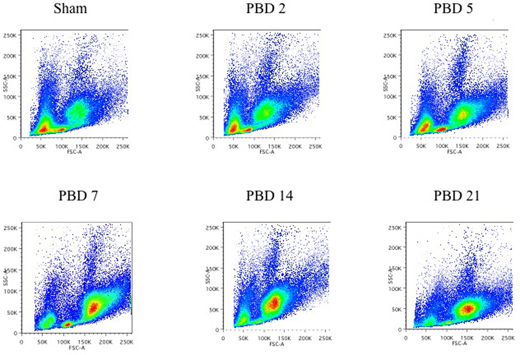
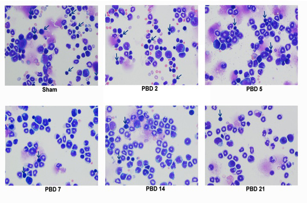
Temporal changes in the erythroid, lymphoid, and myeloid populations following burn injury:
3a) Total bone marrow cells were harvested from sham mice and burn mice on PBD #2, 5, 7, 14, and 21. Flow cytometric analysis of the bone marrow cells on the basis of forward (size) and side (granularity) scatters demonstrated the temporal changes in the erythoid, lymphoid and myeloid populations.
3b) The erythroid, lymphoid, and myeloid populations identified by forward and side scatter were separated by FACS sorting and stained with Wright-Giemsa at various times after burn injury (magnification of 200X). The arrows indicate erythroid progenitors.