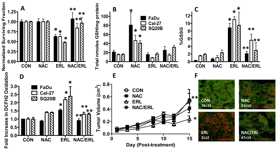
Figure 3. Effect of N-acetyl-cysteine (NAC) on ERL-induced cytotoxicity and oxidative stress.
(A–D) FaDu, Cal-27 and SQ20B cells were treated with 10 µM ERL for 24 hours with or without treatment with 20 mM NAC for 1 hour before and during ERL exposure, then analyzed for clonogenic survival (A), total glutathione (GSH, B), percentage glutathione disulfide (%GSSG, C) and DCFH oxidation (D). Error bars represent ± 1SD of N = 3–4 experiments. (E) Athymic (nu/nu) mice bearing FaDu xenograft tumors were treated with 0.3 g/kg NAC i.p. and/or 12.5 mg/kg ERL p.o. daily for 2 weeks. Control mice received water p.o. daily for 2 weeks. Data points represent the average values for 6 mice. (F) Immunohistochemical analysis of pEGFR expression (green) in tumors. Tumors were counterstained with ToPro3 (red). Red line shown (in the bottom right corner of each image) represents a magnification scale bar of 100 microns. Values shown (in the bottom left corner of each image) represent quantification of pEGFR fluorescence intensity using image analysis and recognition software, Image J and averaged for 3 animals/group for each treatment group. *, p< 0.05 versus control. **, p<0.05 versus ERL.

