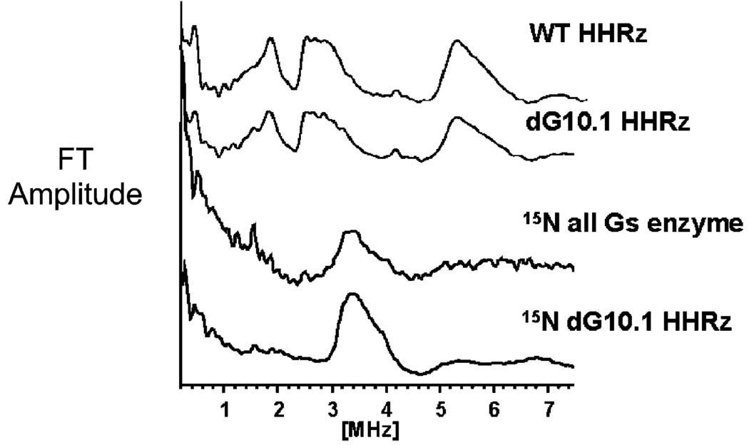
Figure 4.
14/15N three-pulse ESEEM spectra of Mn2+ bound to the hammerhead ribozyme. Wild type and dG10.1 HHRz show similar 14N (I = 1) ESEEM Fourier transform traces, consistent with Mn–guanine coordination. The “15N all G’s enzyme” HHRz, in which all guanines on the enzyme strand are labeled with 15N, shows an ESEEM pattern consistent with Mn–15N (I = ½) coordination.22 A nearly identical spectrum is observed with specific 15N labeling of G10.1, identifying G10.1 as the Mn2+ ligand. Experiment conditions: magnetic field strength 3600 G, tau = 192 ns, η/2 pulse length 15 ns, microwave power 3.2 W.

