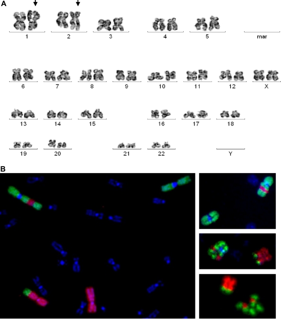
Figure 1.
Cytogenetic aberrations in MSC N0806. (A) G-banding image demonstrates translocation of chromosomes 1 and 2 (arrows). (B) FISH with library probes for whole chromosomes 1 and 2 confirms that a fragment of 1p (green) is translocated to 2q (red) and part of 2q (red) is inserted into 1p (green).

