
Figure 7.
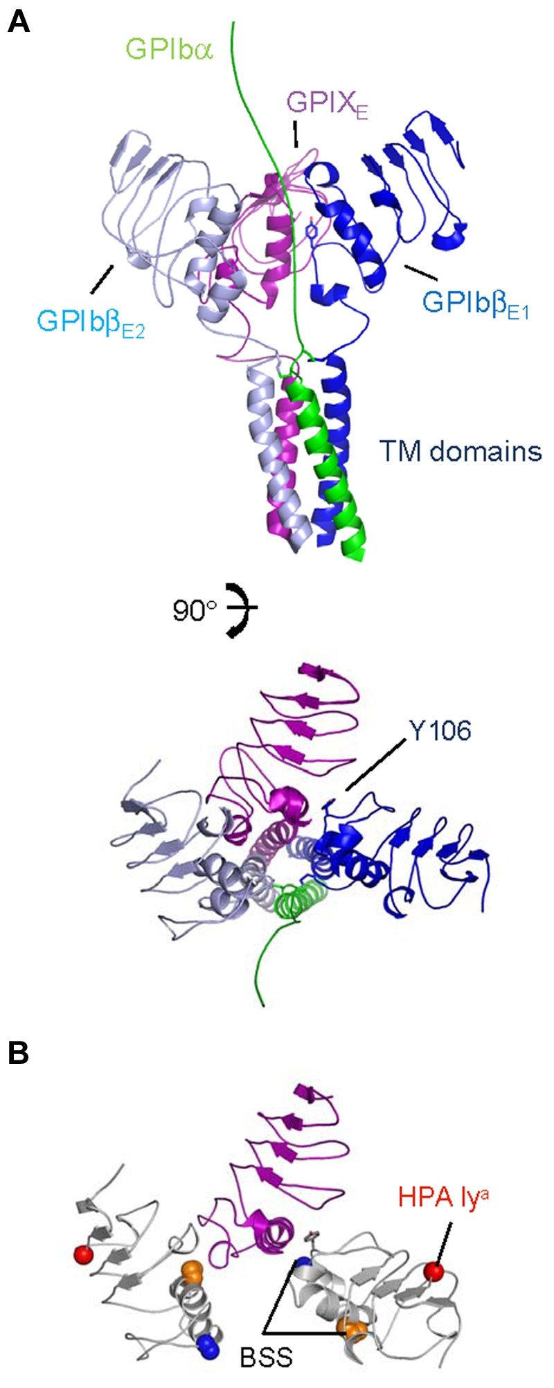
A schematic model for the membrane-proximal portion of the GPIb-IX complex. (A) Cartoon diagrams related by a 90-degree rotation showing the GPIbα chain TM domain (in green) extending toward the N-terminus (top), GPIbβ ectodomain and TM domain (in sky blue and dark blue), and GPIX ectodomain and TM domain (in purple). Tyr106 from GPIbβ1 is shown as stick along with interchain disulphides. (B) Same view as panel A, except without TM domains. GPIbβ is colored white, showing residues affected by BSS and human platelet-specific alloantigen (HPA) mutations. Side chains of GPIbβ residues Ala108 (blue), Pro74 (orange), and Gly15 (red) are shown in space-filling mode; Tyr106 is shown as stick (gray).