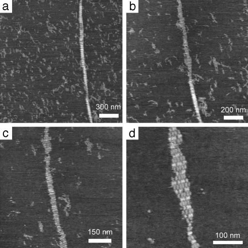
Fig. 3.
A series of AFM images captured by repeatedly reimaging and zooming in on the same nanotube, which appears mostly tube-like in a, with increasing wear-and-tear through b and c until a section of unfolded tube becomes a single-layer flat lattice (d) displaying stripes composed of lighter (higher) B tiles and darker (shorter) A tiles.

