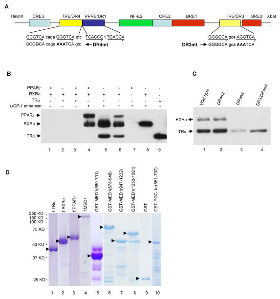
Figure 2. In Vitro Recruitment of TRα, RXRα and PPARγ to the UCP-1 enhancer.
(A). Schematic representation of the UCP-1 enhancer showing the positions of the different elements and the mutated DR4 and DR3 elements.
(B). Independent and joint recruitment of purified PPARγ, RXRα and TRα to the UCP-1 enhancer. Standard immobilized template recruitment assays with proteins added as indicated were carried out as described in Experimental Procedures. Bound proteins in B and C were detected by immunoblot.
(C). Binding of TRα-RXRα to the DR3 enhancer element. Recruitment assays were carried out with TRα, RXRα and either wild type or mutant enhancers (shown in A) as described in Experimental Procedures.
(D). Purified Flag-tagged proteins from sf9 cells (lanes 1–4) and GST-fusion proteins from bacteria (lanes 5–10) analyzed by SDS-PAGE with Coomassie blue staining. Solid arrowheads indicate the corresponding full length proteins.

