
Figure 2.
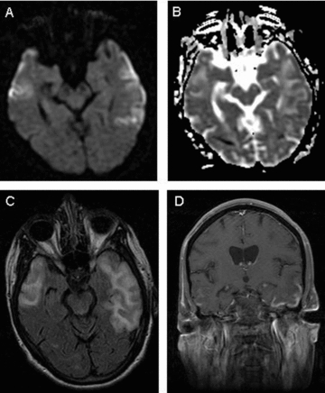
MRI brain. (A) Diffusion weighted image-revealing restriction of diffusion in gyriform pattern in bilateral temporal lobes, worse on the left, confirmed with apparent diffusion coefficient map (B). (C) FLAIR image showing hyperintesities in bilateral lateral temporal lobes. (D) Gyriform pattern of enhancement on postcontrast T1 image.