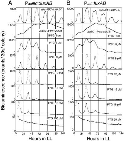
Fig. 2.
Quantitative assay of IPTG dose-dependent restoration of the circadian rhythm in a kaiBC– strain. A kaiABC gene cluster was introduced back into a kaiABC-deleted strain (ΔkaiABC+kaiABC, top trace). Ptrc::kaiCB was introduced into a kaiBC-deleted strain (kaiBC– +Ptrc::kaiCB, lower traces). PkaiBC::luxAB (A) and Ptrc::luxAB (B) reporter cells are shown. Assays for bioluminescence rhythms and presentation of data are the same as described in Fig. 1. Note that the scale for the bioluminescence profile differs among panels. In the lower 12 panels, the concentrations of IPTG indicated in the figure were administered in the solid medium 48–60 h before experimental LL.

