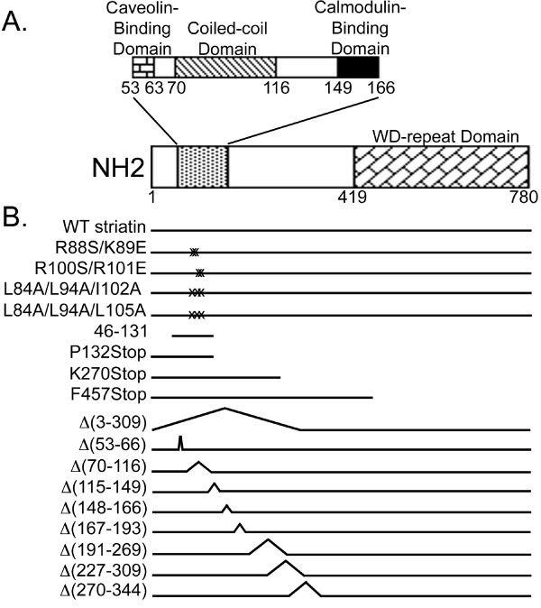
Figure 1.
Schematic of the structures of human wild-type and mutant striatin proteins. (A) Schematic of wild-type striatin, drawn to scale, showing the locations of its previously published protein-interaction domains. White boxes represent regions of unknown function. (B) Stick diagrams of wild-type and mutant striatins used in this study. Point mutations are denoted by X's, and deletions are shown as peaks in the stick diagrams or absence of a line. Positions of mutants are noted relative to wild-type striatin domains in panel A.

