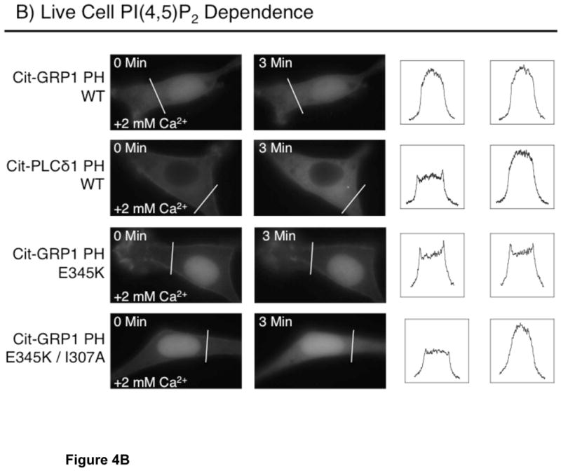
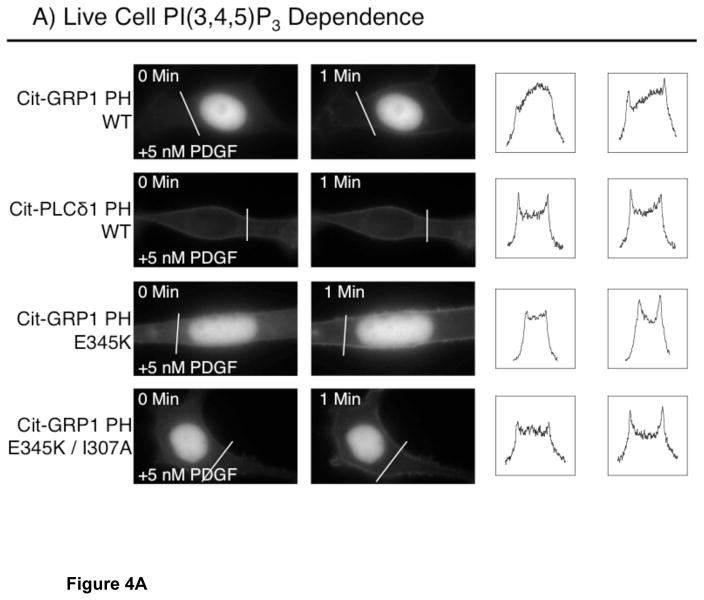
FIGURE 4.

PIP lipid dependence of plasma membrane targeting in NIH 3T3 cells. (A) Dependence of membrane targeting on PI(3,4,5)P3. Shown are the zero time and one minute images for the indicated citrine-coupled PH domains. Serum-starved NIH 3T3 cells were stimulated with 5 nM PDGF at time zero and imaged at multiple timepoints (see Methods). (B) Dependence of membrane targeting on PI(4,5)P2 for the indicated citrine-coupled PH domains. Shown are the zero time and three minute images for the indicated citrine-coupled PH domains. Serum-starved NIH 3T3 cells were incubated in 20 μM ionomycin in Ca2+-free buffer, then stimulated at time zero with 2 mM extracellular Ca2+ to trigger cytoplasmic Ca2+ influx and Ca2+-activated PI(4,5)P2 hydrolysis (45) with imaging at multiple timepoints (see Methods). Each set of images are representative examples of responses observed for 12 or more cells. To assist visualization of the protein movements between the plasma membrane and cytoplasm, fluorescence intensities for pixels defined by the indicated line across each cell are plotted for both before (left box) and after (right box) treatment.

