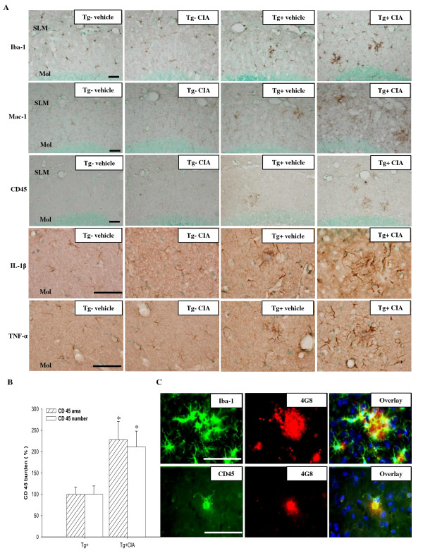
Figure 3.
CIA enhances activation of microglia/macrophages in APP/PS1 mice. (A) Photomicrographs of the dentate gyrus molecular layer immunolabeled with antibodies against Iba-1, Mac-1, CD45, IL-1β, and TNF-α 4 months after injection of 2-month-old wild-type (Tg-) and APP/PS1 mice (Tg+) with vehicle or CIA. SLM, stratum lacunosum molecular; Mol, dentate gyrus molecular layer. Scale bar = 50 μm. (B) Activation of microglia/macrophages was analyzed by measuring the area and the number of CD45-immunoreactivity in the hippocampal and cortical regions (Tg+ vehicle, n = 3; Tg+ CIA, n = 4). (C) Fluorescence photomicrographs showing Iba-1 and CD45 signal around 4G8-positive amyloid plaques in the cortex of CIA-treated APP/PS1 mice. Top row, triple staining with Iba-1, 4G8, and DAPI (a nuclear marker); bottom row, triple staining with CD45, 4G8, and DAPI. Scale bar = 200 μm.

