Figure 5.
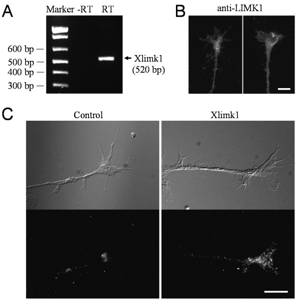
Detection of Xenopus limk1 in Xenopus neurons. (A) RT-PCR detection of Xlimk1 mRNA from RNA samples extracted from Stage 20-22 Xenopus neural tube tissues using specific primers. RNA samples were processed without (-RT) and with reverse transcriptase (RT). (B) Representative fluorescence images of cultured Xenopus growth cones labeled using a specific antibody against LIMK1. (C) Detection of Xlimk1 mRNA in Xenopus growth cones by fluorescence in situ hybridization. Top panels are the differential interference contrast (DIC) images of the growth cones. Bottom panels are the FISH images of Xenopus growth cones labeled with digoxigenin-conjugated probes (three probes, ~50 nt each) that are specifically complementary to different parts of the coding region of Xlimk1 mRNA. The reverse probes were used as the control. Scale: 10 μm.

