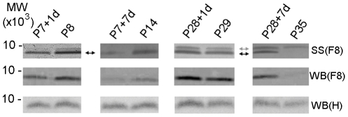
Figure 3. Silver staining and western blots of ubiquitin.
Ubiquitin identified in silver stained (SS) gels and in western blots (WB) of Fraction 8 (F8) and in un-fractionated homogenate (H) of the cord segment caudal to the injury site. Ubiquitin was identified by mass spectrometry only in Fraction 8 (pH 7.06–7.64). Silver stained gels showed that in the P7-injured groups, a single band (black arrow) was detected. One day after injury (P7+1d) ubiquitin decreased compared to age-matched controls of P8 and the decrease was more pronounced seven days after injury (P7+7d). In the P28-injury groups, ubiquitin increased both 1 day (P28+1d) and especially 7 days (P28+7d) after injury compared to age-matched controls (P29 and P35 respectively). An additional band approximately 1 kDa higher (gray arrow) was also detected at these older ages. Western blots of Fraction 8 confirmed changes observed by silver staining. However, the antibody used was only able to detect the lower band and did not recognize the additional band observed in the P28-injury groups. Western blotting of the whole un-fractionated homogenate, (WB) H confirmed changes observed in silver stained gels although the differences were less pronounced. MW: Molecular weight marker (MW×103).

