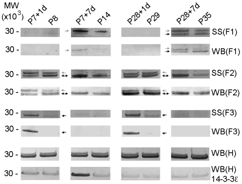
Figure 4. Silver staining and western blots of 14-3-3 protein family.
Family of 14-3-3 proteins identified from silver stained gels (SS) and western blots (WB) of Fractions 1, 2 and 3 (F1, F2 and F3) and western blots of un-fractionated homogenate (WB) H of spinal cord segment caudal to the site of injury. Two subtypes, 14-3-3 epsilon and gamma were identified in silver stained gels and detected by mass spectrometry in F1, F2 and F3, pH 3.00–3.58, pH 3.58–4.16 and pH 4.16–4.76, respectively. 14-3-3 epsilon subtype was identified as the higher band (grey arrow) whilst 14-3-3 gamma subtype was identified as the lower band (black arrow). Results showed that 14-3-3 epsilon increased 7 days after injury at both ages (P7+7d and P28+7d) compared to age-matched controls (P14 and P35) in F1, while in F2 it increased 1 day after injury at P7 only (P7+1d). 14-3-3 gamma (lower band) was predominantly expressed in F2 at all ages but was additionally detectable in F1 at P35 and F3 at P8 and P29. In the P7-injured group, 14-3-3 gamma was initially down-regulated at 1 day after injury but was up-regulated 7 days after in F2. However in F3, it was up-regulated 1 day after injury but was not detectable at P14. In P28-injured group it showed differential expression in all fractions where it was down-regulated in F2 but was up-regulated in F3 one day after spinal injury. Seven days after injury, it was down-regulated in F1 but up-regulated in F2. Silver stained gels of F1-F3 were confirmed using pan-14-3-3 antibody and western blotting (WB). Western blotting of un-fractionated homogenates (H), which contain all 14-3-3 subtypes, with the pan-14-3-3 antibody showed no detectable overall changes in expression levels in the samples from spinal cords after injury compared to age-matched controls. Western blotting of un-fractionated homogenates with anti-14-3-3 epsilon showed that there was an increase in 14-3-3 epsilon expression at P7+1d and P7+7d compared to age-matched control. No changes were observed in P28-injured group compared to age-matched control. MW: Molecular weight marker (MW×103).

