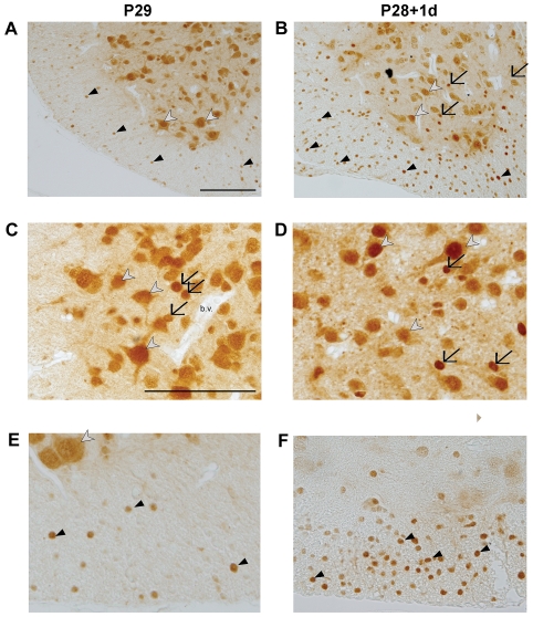
Figure 7. Immunocytochemical detection of ubiquitin in the spinal cord at P28+1d and P29.
Immunocytochemical detection of ubiquitin at P29 (left panel) and P28+1d (right panel) of caudal section of the cord (A and B), grey matter (C and D) and white matter (E and F). In sections from both control and injured cords, ubiquitin was localized in nuclei of neurons (white arrowheads) and glial cells (black arrows) in the grey matter and oligodendrocytes in the white matter (black arrowheads). In addition, ubiquitin was detected in the cytoplasm of neurons. Identification of cell types was based on the morphology of the cell and comparison with rat tissue where cell-specific markers such as olig2 for oligodendrocytes and NeuN for neurons are available. Increased staining of oligodendrocytes (white matter) and glial cells (grey matter) was observed one day after injury as indicated by black arrows and arrowheads in (B, D, F). Similar pattern was visible 7 days after injury (not illustrated). Blood vessels (b.v. in C) were negative in all sections. All scale bars are 100 µm. Scale bar in A applies also to B. Scale bar in C applies to figures in C–F.

