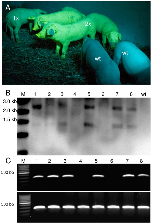
Figure 5. Segregation of Venus transposon in F1 offspring.
A) F1-offspring viewed under specific excitation of Venus. Under the recording conditions, Venus fluorescence is displayed as green-yellow color, exemplarily some animals are labelled with their copy number of the transposon. Note that the copy numbers of the transposon correlated directly with the fluorescence intensity. The non-transgenic littermates (wt) appear bluish due to reflected and scattered excitation light B) Southern blot analysis of one litter. Lanes 1-3, 5, 7 and 8 represent transposon transgenic piglets with specific hybridization signals. Lanes 4 and 6 represent non-transgenic littermates. M, molecular size marker. C) PCR genotyping for the presence of the Venus transgene. As positive control, a PCR for polyA polymerase (PolyA) was included; wt, wild type sample.

