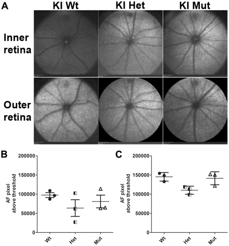
Figure 5. Fundus autofluorescence (AF) in C1qtnf5 wild-type and Ser163Arg knock-in mice.
A) Representative fundus images of inner and outer retina obtained by AF-SLO imaging from wild-type, heterozygous and homozygous mutant C1qtnf5 S163 KI mice at 16–18 months of age. No obvious difference in clearly demarcated autofluorescent areas or punctuated pattterns in the fundus images were observed in any of the three gentotypes. Wt: wild-type mice, Het: heterozygous KI mice, Mut: homozygous KI mice. B,C) Semiquantitative analysis of the inner (B) and outer (C) retinal background autofluorescence. The mean background autofluorescence (AF-pixel above threshold) for the right and left eye per animal is shown for each genotype (n = 3 animals per genotype). No significant difference in background autofluorescence in the inner retina (B, Kruskal-Wallis with Dunn's multiple comparison test, p = 0.393) or in the outer retina (C, Kruskal-Wallis with Dunn's multiple comparison test, p = 0.067) was observed between any of the three gentoytpes.

