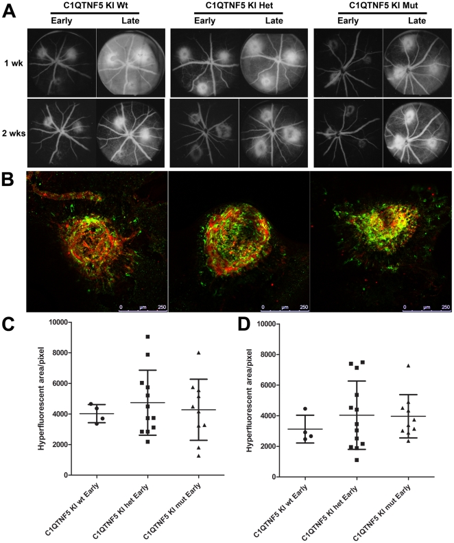
Figure 7. Laser-induced choroidal neovascularization in wild-type and C1qtnf5 Ser163 Arg knock-in mice.
A, Fundus images from the early and late phase of in vivo fundus fluorescein angiography at 1 and 2 weeks after photocoagulation/laser induced choroidal neovascularization (CNV) in 15–16 month old wild-type and C1qtnf5 KI mice. Wt: wild-type mice, Het: heterozygous KI mice, Mut: homozygous KI mice. B, The microglia in laser lesions on RPE flat mounts resulting from CNV laser treated C1qtnf5 KI mice after 2 weeks were labelled with anti-Iba1 (green) and co-labelled with anti-lectin (blood vessels) antibodies. Scale bar, 250 µm. C,D, Quantitative analysis of area of hyperfluorescence in the fundus images from 1 week old animals (C) and 2 week old animals (D) as a measure for CNV lesion size. No significant difference between the hyperfluorescent areas were observed at 1 week (Kruskal-Wallis with Dunn's mutliple comparison test, p = 0.911) or 2 weeks (Kruskal-Wallis with Dunn's mutliple comparison test, p = 0.638). The number of animals for both time points were n (wt) = 4, n (het) = 12, n(mut) = 10.

