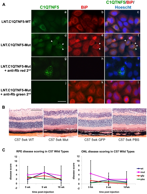
Figure 8. Effects of Lentiviral expression of wild-type or Ser163Arg mutant C1qtnf5 in mouse retinas.
(A) HeLa cells were infected with lentivirus contain wild-type C1QTNF5 (LNT.C1QTNF5-WT) or Ser163Arg mutant C1QTNF5 (LNT.C1QTNF5-Mut). At 72 hr post-infection, cells were stained with anti-CTRP5 (green) and the endoplasmic reticulum (ER) marker anti-BiP (red) antibodies. Cells infected with LNT.CTRP5 or LNT. CTRP5-Mut both showed intracellular staining surrounding the nucleus (a, d) suggesting that both proteins enter the secretory pathway. While the staining on LNT-CTRP5 infected cells is rather diffuse (a), the LNT-CTRP5-Mut showed punctate staining (d) that co-localised with the ER marker BiP (e, f, shown by white arrow heads) suggesting that the mutant form of the protein is retained in the cell and cannot exit the ER correctly. Controls showed that there was no contribution to the fluorescent signals from the respective anti-rabbit secondary antibodies (g, h, i, j, k, l). Scale bar = 20 µm. (B) Haematoxylin and eosin stained retinal sections from C57BL/6 mouse eyes at 5 weeks after subretinal injection of either LNT.C1QTNF5-WT, LNT.C1QTNF5-Mut, LNT.GFP or PBS. No obvious differences could be observed between the four groups despite some injection related trauma (not shown here). (C) RPE and outer nuclear layer (ONL) disease scoring was assessed on randomized images and showed no significant difference between the any of the four treated groups, n = 4 per group (One-way ANOVA, all p-values p>0.05). Scale bar = 100 µm.

