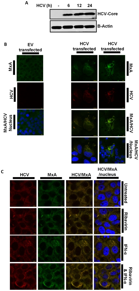
Figure 4. HCV core co-localises with MxA.
(A) Immunoblot of lysates from Huh7 cells transfected with HCV-DNA construct for 0, 6, 12 and 24 h, probed with HCV core and β-Actin (n = 3). (B) Confocal micrograph of Huh7s transfected with EV or HCV-DNA construct (n = 3). (C) Huh7s transfected with HCV-DNA construct and treated with IFN-α for 2 h, Ribavirin for 2 h or both for 2 h (n = 4). Confocal micrographs show MxA, HCV core and the nucleus, labelled with Alexa 488, Alexa 568 and DAPI, respectively. Antibody staining is indicated along the side of images Bar, 10 µm (n = 3).

