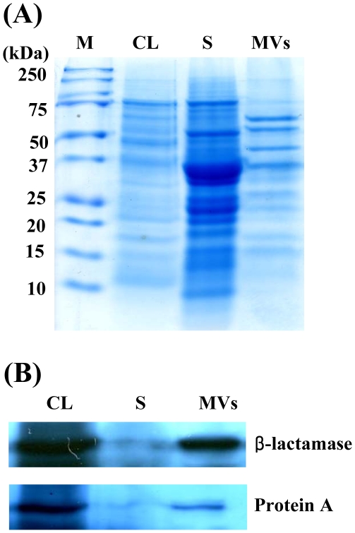
Figure 2. Proteins identified in the S. aureus-derived MVs.
SDS-PAGE of proteins packaged in the MVs from S. aureus 06ST1048 (A) and its Western blot analysis (B). The samples were separated on 12% SDS-PAGE and immunoblotted with anti-protein A and anti-β-lactamases antibodies. Lanes M, molecular weight maker; CL, bacterial cell lysate fraction; S, supernatant fraction; and MVs, membrane-derived vesicle fraction.

