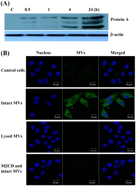
Figure 3. Delivery of protein A packaged in the S. aureus MVs to host cells.
(A) Delivery of protein A to host cells via the MVs. HEp-2 cells were treated with S. aureus MVs (20 µg/ml of protein concentrations) for the indicated times. Cell lysates were separated on 12% SDS-PAGE, transferred to membranes, and immunoblotted with anti-protein A and β-actin antibodies. Both full-length and degraded forms of protein A were appeared. (B) HEp-2 cells were treated with intact or lysed MVs (20 µg/ml of protein concentrations) for 24 h. HEp-2 cells were pretreated with 10 mM MβCD for 45 min at 37°C. The cells were fixed, permeabilized, and stained with a rabbit anti-protein A antibody, followed by Alexa Fluor® 488-conjugated rabbit IgG (green). DAPI was used to stain the nuclei (blue). The analytical sectioning was performed from the top to the bottom of the cells. The figure represents all projection of sections in one picture.

