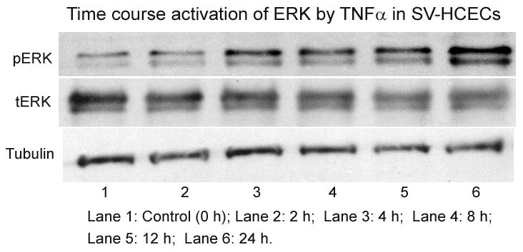
Figure 2. Time-course study of ERK activation in SV-HCECs by cytokine.

Cells were incubated with and TNFα (100 ng/ml) at different time period from 0 h (control) to 24 h. Cells were collected and protein was dissolved in Lemmili’s buffer. ERK activation was measured using Western blot as described in the text.
