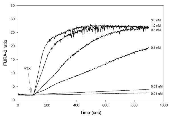
Figure 1.
Effect of maitotoxin (MTX) on fura-2 fluorescence in bovine aortic endothelial cells (BAECs). Fura-2-loaded BAECs were suspended in HBS and the fluorescence ratio recorded as a function of time as described in Materials and Methods. Six traces are shown superimposed. At time 100 sec, MTX was added during the individual recordings at the final concentration indicated to the right of each trace. Results shown are representative of 11 individual experiments.

