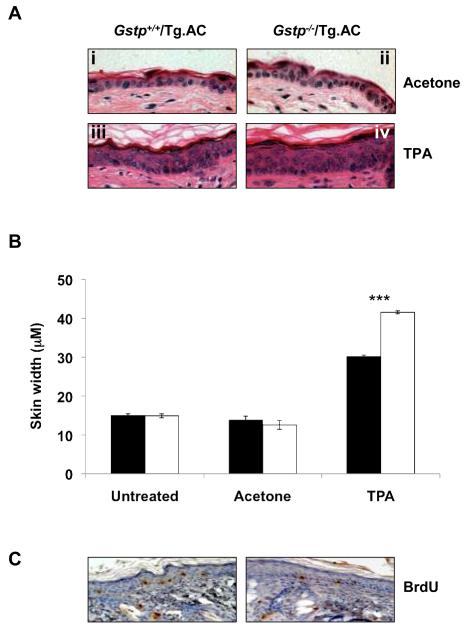
Figure 3. Increased skin thickness, but no change in BrdU labelling, in Gstp−/−/Tg.AC mice treated with TPA for 6 weeks.
(a) Increase in skin thickness relative to acetone controls in both Gstp+/+/Tg.AC (compare i and iii) and Gstp−/−/Tg.AC mice (compare ii and iv) following TPA treatment, whilst comparison of iii and iv clearly shows the increased thickness of the epidermal layer in Gstp−/−/Tg.AC mice relative to Gstp+/+/Tg.AC mice. *** p<0.001; 6 fields/slide, 5 measurements/field; n=3, (b) Skin thickness in Gstp+/+/Tg.AC (black bars) and Gstp−/−/Tg.AC (white bars) mice following TPA treatment, (c) No difference in BrdU-labelled cells between Gstp+/+/Tg.AC mice and Gstp−/−/Tg.AC mice. Sections shown are representative of each genotype (3 fields/slide, n= 3 for each genotype).

