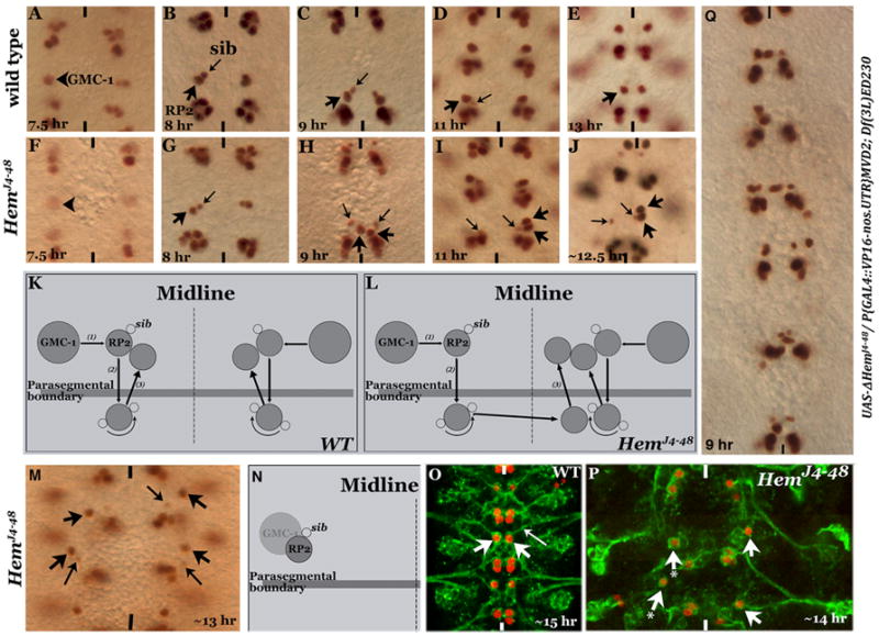
Figure 2. The RP2 defect in Hem mutants is due to aberrant migration.

Embryos in panels A-J, M and Q are stained with anti-Eve, and in panels O and P are stained with anti-Eve and 22C10 antibodies. Anterior end is up, midline is marked by vertical lines. Panels A-E: Wild type embryos showing the 3-step RP2/sib migration. Panels F-J: Hem mutant embryos at the corresponding developmental stages. Panels K and L: Line drawings depicting RP2/sib migration in wild type and Hem mutants. Panel M: RP2 fails to migrate and is located at the position of its parent GMC-1. Panel N: Line drawing illustrating the migration defect. Panel O: Wild type embryo showing axon projection (22C10 positive) from an RP2, fasciculating with ISN. Panel P: Hem mutant embryo, RP2 fails to migrate from its location of formation; this RP2 has no discernible axonal projection (arrow-with-star). Panel Q: Hem-deficiency embryo with truncated Hem expressed from a UAS-DHem transgene.
