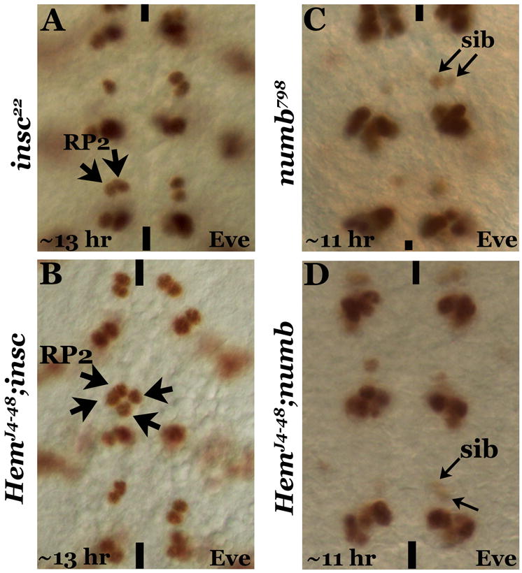
Figure 3. The migration defect is specific to RP2 but not its sib.

Embryos are stained with anti-Eve, anterior end is up, midline is marked by vertical lines. Panel A: insc mutant with the duplication of RP2. Panel B: HemJ4-48; insc double mutant showing four RP2s on one hemisegment and none on the contralateral hemisegment. Panel C: numb mutant showing the duplication of sib. Panel D: HemJ4-48; numb double mutant showing the numb-phenotype without any sib migration across the midline.
